
Necrotising fasciitis is a rare but potentially lethal infection. Abdominal wall necrotising fasciitis that was associated with mortality.

Pin On Exercise
In the largest series of 53 patients only two patients had PF lesion arising from the flank.

. Abstract We report three cases of abdominal wall necrotizing fasciitis that occurred as a result of leakage from displaced percutaneous endoscopic gastrostomy tubes. Only few hundred cases have been reported in the literature. The author reports here the case of a woman with necrotizing fasciitis of the abdominal wall and the course and methods of treatment.
Necrotizing fasciitis is a deep infection of the subcutaneous tissue that results in progressive destruction of fascia and fat. The abdominal wall defect may shrink during the last trimester referred to as closing gastroschisis this can lead to compression of the herniated viscera resulting in intestinal obstruction andor mesenteric ischemia. Our case illustrated the rare complication related to the insertion of PEG tube.
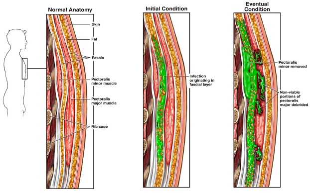
Nodular fasciitis is a rare noncancerous tumor. When necrotising fasciitis appears in the abdominal wall the tissue must be removed with a surgical procedure called debridement. Find read and cite all the research you.
Although immunodeficiency is a risk factor for NF there is only one reported case of NF in AIDS involving the cervical region. Hence the mortality rate is high median mortality 322. Dislodgement of the gastrostomy tube is implicated in most cases as in this case and allows infection by gas-producing organisms.
Two comorbidity factors extreme obesity diabetes and the late diagnosis of necrotizing fasciitis the latter masked by celullitis and phlegmona of the abdominal wall resulted in overdue adequate surgical. It can affect soft tissues across the body including the abdominal wall. An epigastric hernia occurs when a weakened area in the abdominal wall allows a bit of fat to push through.
Patients underwent extensive operative excisions of their abdominal walls down to their posterior fascia. Authors Matthew Jacob Smith 1 2 Ranah Lim 1 2 Mark Damien Muhlmann 1 2 Affiliations 1 Department of General Surgery Prince of Wales Hospital Sydney. The infection progresses rapidly and septic shock may ensue.
Spigelian hernias can also occur in a traumatic event and rarely there are trans-diaphragmatic intercostal hernias. Computed Topography CT scan confirmed abdominal wall necrotising fasciitis complicated with hyperosmolar hyperglycaemia state HHS and later succumbed after 48 hours of admission. Epigastric hernias are typically small.
Mortality rate is high and has not changed since it was first described by Meleny. Nodular fasciitis mimics malignant cancerous tumors which makes it a challenge to diagnose. PDF We report a case of non-necrotizing abdominal wall fasciitis as a post-operative complication of percutaneous endoscopic gastrostomy insertion.
Protects the abdominal viscera from injury. Abdominal wall necrotising fasciitis with colonic fistula ANZ J Surg. Morel-Lavallée lesions affect mostly the subcutaneous tissue.
Abdominal wall necrotizing fasciitis is a rare complication of percutaneous gastrostomy with an estimated incidence of 1 in 129 gastrostomy procedures 2. This is referred to as closed gastroschisis. Perforation after bowel strangulation causes sepsis which may cause peritonitis or spread to the soft tissue of the anterior abdominal wall.
Online ahead of print. Abdominal wall pain can be thought of as one category of myofascial pain. Necrotizing fasciitis NF is a severe rare potentially lethal soft tissue infection that develops in the scrotum and perineum the abdominal wall or the extremities.
In rare cases a complete closure of the abdominal wall at birth can be observed. Forms a firm yet flexible boundary which keeps the abdominal viscera in the abdominal cavity and assists the viscera in maintaining their anatomical position against gravity. Traumatic abdominal wall hernias usually occur at various weak spots of the abdominal wall most frequent are lumbar hernias affecting the inferior lumbar triangle.
They occur in the middle of the belly in the area between the belly button and the breastbone. A failure to do so will result in fatal complications. We present the case of a 59-year-old man with recurrent groin infections poorly controlled Type 2 diabetes and obesity who developed necrotizing fasciitis of his lower abdominal wall secondary to Actinomyces europaeus.
Some patients develop more than one epigastric hernia at a time. The disease is classified as type I polymicrobial infection type II monomicrobial and type III gas gangrene or clostridial myonecrosis. In this situation muscle or fascial strain can lead to a pain trigger point.
The abdominal wall encloses the abdominal cavity and can be divided into anterolateral and posterior sections. We discuss the clinical course and the value of early identification of the pathogen and specialist microbiologist advice. Proliferative fasciitis PF is a rare pseudosarcomatous lesion arising from the subcutaneous fascia and the fibrous septa.
Necrotizing fasciitis of abdominal wall in AIDS Necrotizing fasciitis NF is an uncommon but potentially lethal soft-tissue infection. Abdominal wall fasciitis as a complication of abdominal wall hernia occurs when there is a delay in management of associated bowel strangulation and subsequent perforation. It can appear in soft tissue anywhere on your body.
This is the first report of such a series.
![]()
Necrotizing Fasciitis Diagnosis Treatment Images Kenhub

Pin On Mind Body Connection

Actual And Potential Perineopelvic Spaces Anatomy Sagittal Section Visceral Peritoneum Vesical Fascia Rectal Fasc Medical Anatomy Anatomy Pelvic Diaphragm

Pin On Knee Pain

Necrotizing Faschiitis With Necrosis Of Skin Vascular Thrombosis And Involvement Of Underlying Mus Subcutaneous Tissue Medical Illustration Wound Care Nursing
Pin Em Being A Nurse

Necrotizing Fasciitis Of The Abdominal Wall Cjs
Necrotizing Fasciitis Diagnoses And Therapy Acep Now

Plantar Fasciitis 3d Medical Vector Illustration On White Background Illustration Spons Plantar Fasciitis Relief Plantar Fasciitis Treatment Plantar Fasciitis

Pin On Core

Abdominal Wall Fascia Sports Physiotherapy Stefan Duell Facebook

Pin On Touchstone Massage Therapy

Pin On Medical Forum From Jama

This Diagram Uses The Torso And Arm To Show How Skeletal Muscles Are Typically Attached To Bones By Tendons Body Anatomy Male Torso Human Anatomy

Pin On Vezbice
![]()
Necrotizing Fasciitis Diagnosis Treatment Images Kenhub

How Determine If You Need Surgery For Plantar Fasciitis Plantar Fasciitis Fascia Stretching Plantar Fascia Tear
:format(jpeg)/images/article/en/clinical-case-necrotising-fasciitis-of-the-anterior-abdominal-wall/39VXCIiT9KP6iqdHHJekHg_Necrotizing_Fasciitis_of_the_Anterior_Abdominal_Wall.png)
Necrotizing Fasciitis Diagnosis Treatment Images Kenhub

Pin On Plantar Fasciitis